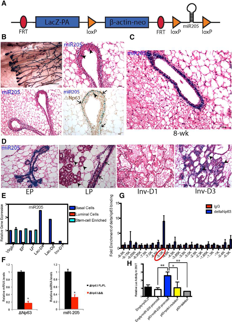
Figure 1.

miR-205 is predominantly expressed in the basal/stem cell enriched population of the postnatal mammary gland. (A): Structure of the targeted miR-205 locus, in which the upstream lacZ fragment can be used to as a reporter of miR-205 expression. (B): MiR-205 is expressed in the outer cap cell layer of terminal end buds (TEBs) as well as the subtending ducts and co-localizes with △Np63 (arrows). There are also miR-205+/△Np63+ cells in a small number of body cells (arrowheads) of TEBs. (C): MiR-205 is expressed in the basal layer of 8-week postnatal mammary gland. (D): MiR-205 expression pattern in early pregnancy (EP: day 6 of pregnancy), late pregnancy (LP: day 18 of pregnancy), and involution day 1 and day 3. (E): MiR-205 expression pattern across different stages of development. (F): miR-205 expression displays remarkably decrease in the primary culture of Ad-cre treated △Np63fl/fl mammary epithelial cells (MECs). (G): The −5 Kb region upstream of miR-205 gene is bound by △Np63 as shown by ChIP-quantitative polymerase chain reaction (qPCR). The 20 primer sets were used to amplify the −10 Kb region upstream of miR-205 with ~500 bp intervals. (H): The miR-205 promoter region containing p63 BS was cloned into pGL3 reporter and the luciferase activity was measured with WT and mutated p63 BS. *p < .05; **p < .01 by unpaired Student’s t tests. Scale bars: (B) left panel 10 um; (B) right panel, (C), and (D) 20 um.
