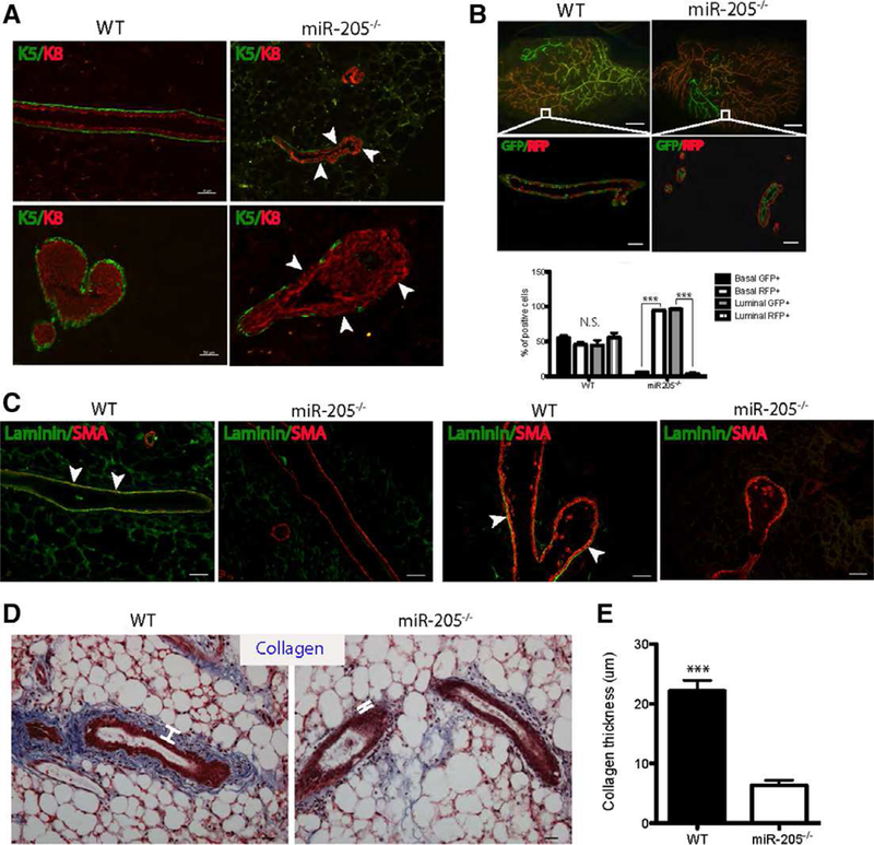
Figure 3.

Ablation of miR-205 leads to alterations in the basal cells, extracellular matrix and stroma. (A): Immunofluorescence (IF) for K5 and K8 demonstrating the significant loss of K5+ basal cells (arrowheads) in miR-205−/− ducts (upper panel) and terminal end buds (TEBs) (lower panel) compared to that of WT. (B): Yellow WT and miR-205−/− outgrowths (white squares, generated from 100,000 MECs transplantation) were sectioned and stained with GFP and RFP. The quantification of the number of GFP/RFP+cells showed that in miR-205−/− glands, the basal compartment was mainly comprised of RFP+ cells, whereas the luminal compartment was mainly comprised of GFP+cells. (C): IF for laminin and αSMA demonstrating the loss of basement membrane in both the ducts and TEBs in miR-205−/− outgrowth compared to that of WT. (D): Trichrome staining depicting significant loss of collagen in miR-205−/− outgrowths compared to WT. White lines measured the thickness of collagen layers. (E): Collagen thickness in miR-205−/− outgrowths was significantly decreased compared to WT. Data shown in (B) and (E) are mean ± SD from three independent sections. ***p < .001 by unpaired Student’s t tests. Scale bars: (A) and (C) 50 um; lower panel in (B) 50 um; upper panel in (B) 20 um; (D) 20 um.
