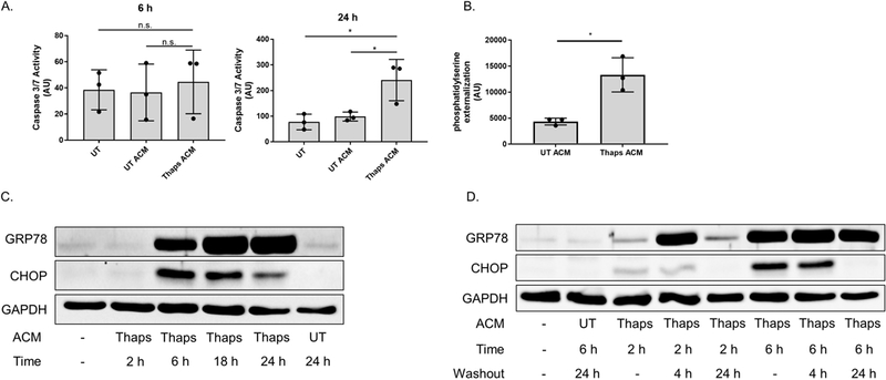
Figure 5. Mild exposure to the stress factor(s) promotes an adaptive state in neurons.

(A) Murine HT-22 hippocampal neurons were stimulated with control or ER stressed astrocyte conditioned media (UT or Thaps ACM) for 6 or 24 h. Caspase-3 activity was assessed by measuring the fluorescence intensity of the cleaved capase-3 substrate Ac-DEVD-AMC. Caspase-3 activity was expressed as arbitrary fluorescence units (AU). N = 3, *P ≤ 0.05 determined by one-way ANOVA with Tukey’s multiple comparisons test. (B) HT-22 neurons were treated with astrocyte conditioned media (UT or Thaps ACM) for 24 h followed by measuring phosphatidylserine externalization using a split luciferase coupled to Annexin V (Real-time Glo), data are expressed as arbitrary luminescent units (AU). N = 3 independent cell culture preparations, *P ≤ 0.05 determined by Student’s T test. (C) HT-22 neurons incubated with 25% control or ER stressed ACM for the indicated time points. GRP78 and CHOP protein expression was examined using immunoblot analysis. (D) HT-22 neurons incubated with 25% control or ER stressed ACM for 2 or 6 h, followed with a 4 or 24 h washout period. GRP78 and CHOP protein expression was examined using immunoblot analysis. GAPDH was used as the loading control. UT, untreated; UTA, control ACM; TA, Thaps ACM.
