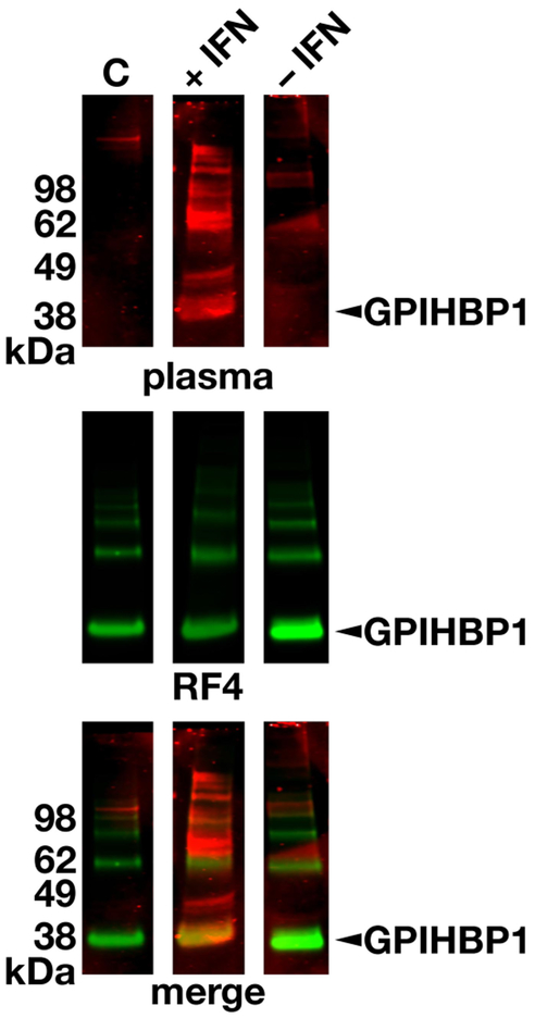
Figure 5.
Detecting GPIHBP1 autoantibodies by western blotting. Proteins in the conditioned medium of human GPIHBP1-expressing Drosophila S2 cells were size-fractionated under nonreducing conditions and then transferred to nitrocellulose membranes. The membranes were incubated with plasma from a normal control subject (C) or the plasma from the multiple sclerosis patient during IFN β1a therapy (+IFN) and after discontinuing IFN therapy (−IFN). Human immunoglobulins were detected with an IRDye680-labeled donkey anti-human IgGs (red, top row). The same membranes were subsequently incubated with an IRDye800-labeled human GPIHBP1-specific antibody RF421 (green, middle row). The bottom row shows the merged images. Arrowhead points to the GPIHBP1 band. GPIHBP1, glycosylphosphatidylinositol-anchored high-density lipoprotein–binding protein 1; IFN, interferon.

