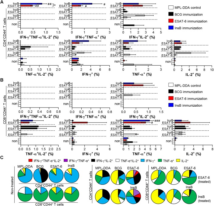
FIGURE 2.
Induction of antigen-specific multifunctional T cells in the lung of ESAT-6- and InsB-immunized mice. At 3 weeks after the last immunization, lung cells from adjuvant (MPL-DDA)-, BCG-, ESAT-6-, or InsB-immunized mice (n = 6 mice/group) were stimulated in vitro with no antigen, with ESAT-6, or with InsB in the presence of GolgiStop for 12 h at 37°C. The percentage of antigen-specific CD3+CD4+CD44+ (A) and CD3+CD8+CD44+ (B) T cells producing IFN-γ, TNF-α, or IL-2 was measured according to the gating strategy shown in Supplementary Figure 1. The frequency of T cells producing each combination of cytokines is shown as the percentage of the specific cell type in the CD3+CD4+CD44+ or CD3+CD8+CD44+ T cell population. The mean ± SDs (n = 6 mice/group) shown are representative of two independent experiments. ∗p < 0.05 (InsB vs. ESAT-6-immunized group) and #p < 0.05, ##p < 0.01, ###p < 0.001 (antigen-immunized groups vs. each antigen-treated MPL-DDA group). To reduce false positives of cytokine-producing populations caused by non-specific staining, fewer than 10 cells in lung and 15 cells in spleen were considered as zero. (C) Pie charts represent the mean frequencies of cells coexpressing IFN-γ, TNF-α, or IL-2.

