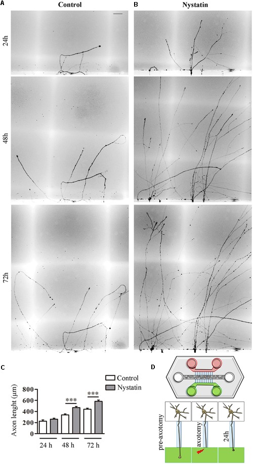
FIGURE 5.

Nystatin improves axon regeneration after axotomy in vitro. Examples of hippocampal axon regrowth at 24, 48, and 72 h after axotomy in control condition (A) and in response to treatment with 2.5 μg/ml Nystatin (B). Axon lengths were measured in each condition. Data represent means ± SEM (C). Schematic diagram of a microfluidic chamber (D). Cell bodies are in the central channel (white), axon growth through microfluidic channels (blue) to both sides (green and red). After axotomy, control medium was applied on one side (red) and Nystatin on the other (green). N = 3 chambers (t-Test in each time point, ∗∗∗p < 0.0001). Scale bar 50 μm.
