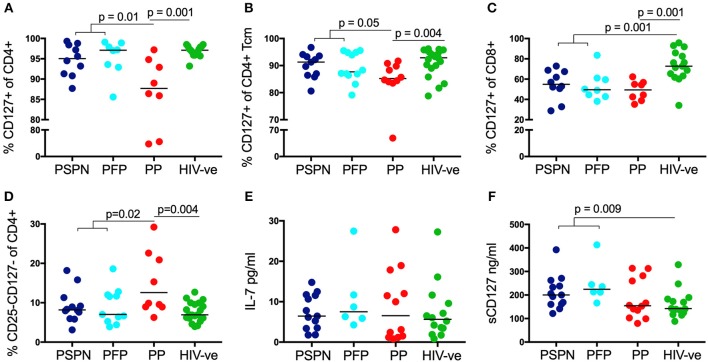
Figure 4.
Increased IL-7/sIL-7 receptor in pediatric slow progressors. Frequency of IL-7R (CD127) expression on CD4 (A) and on central memory CD4 (B) T-cells in PSPN (dark blue; n = 10), PFP (light blue; n = 8), PP (red; n = 8), and uninfected pediatric controls (green; n = 15). (C) Same groups as before but showing frequency of IL-7R expression on CD8 T-cells and (D) frequency of CD4 T-cells double-negative for IL-2R (CD25) and IL-7R (CD127). (E) Ex-vivo plasma levels of IL-7 in pg/ml in PSPN (n = 13, dark blue), PFP (n = 6, light blue), PP (n = 12; red), and pediatric uninfected controls (n = 14; green). (F) Same as in (E) but showing plasma levels of IL-7R (sCD127) in ng/ml in the different groups. For scatterplots, median, and interquartile range are shown. Kruskal-Wallis test was performed and corrected for multiple comparisons.

