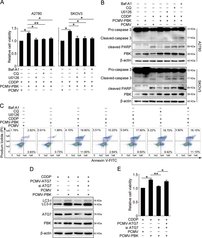
Fig. 5. PBK confers cisplatin resistance through autophagy and ERK/mTOR axis.
Cells were transfected with PCMV or PCMV-PBK for 24 h, then cultured with or without CDDP (5 μg/ml), U0126 (10 μM), CQ (50 μM), and Baf A1 (50 nM) for 24 h. a The CCK8 assay was used to determine the relative cell viability in A2780 and SKOV3 cells. b Western blot analysis of protein levels of cleaved-caspase-3, cleaved PARP, PBK, and β-actin in A2780 and SKOV3 cells. Quantification of relative protein expression levels was shown in Supplementary Figure S9. c The proportion of apoptotic A2780 cells were determined by AnnexinV-FITC/PI staining and flow cytometry. A2780 cells were transfected with PCMV, PCMV-PBK, PCMV-ATG7, or ATG7 siRNA (si ATG7) for 24 h, followed by 5 μg/ml cisplatin treatment for 24 h. d Western blot analysis of protein levels of LC3-I, LC3-II, ATG7, PBK, and β-actin. e The CCK8 assay was performed to detect the relative cell viability (data are mean ± SEM, *p < 0.05, **p < 0.01, n = 3)

