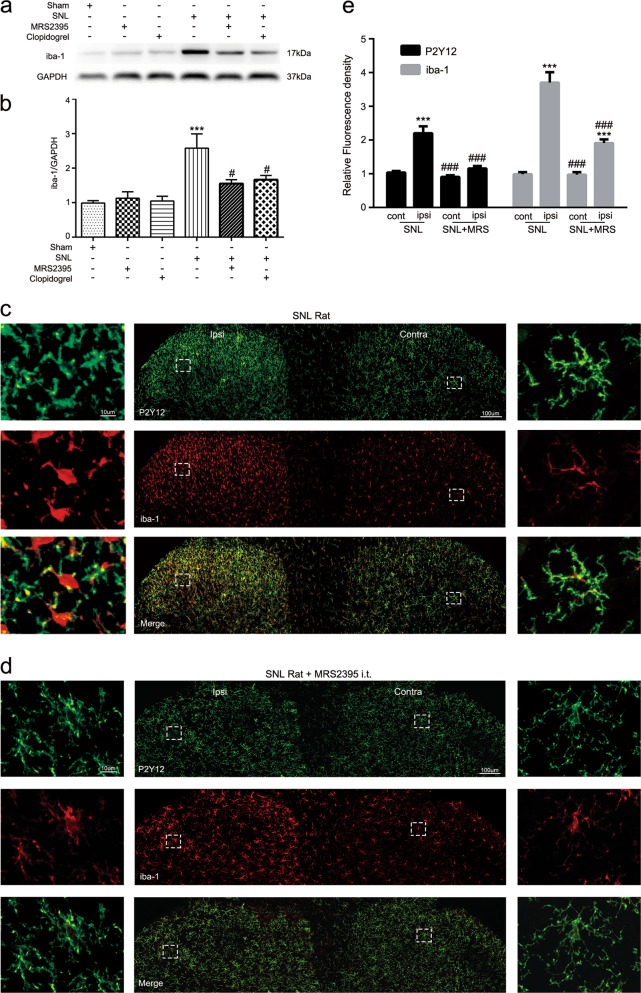
Fig. 4. P2Y12 antagonist attenuated the spinal nerve ligation (SNL)-induced activation of microglia in the spinal dorsal horn of rats.
a Representative immunoblots of iba-1 from total cell lysates of ipsilateral spinal cord tissue harvested 7 days after SNL surgery. GAPDH was used as the loading control. b Densitometric analysis (mean ± SEM, n = 6 rats per group) of iba-1 normalized to the GAPDH loading control. ***p < 0.001 vs. sham group; #p < 0.05 vs. SNL group. c Representative confocal images at low and high magnification of both dorsal horns at postoperative day 7 following SNL in rats. Immunoreactivity of P2Y12 and iba-1 are shown in green and red, repectively. Scale bar: 100 and 10 μm for lower- and higher-magnification images, respectively. d Representative confocal images of MRS2395-treated rats after SNL at low and high magnification. High-magnification images showed that after injection of MRS2395, microglia morphology on the ipsilateral side transformed from an activated state (with swollen cell bodies and retracted processes) to a nonactivated state (with small cell bodies and more ramified processes). e Quantified immunoreactivity of P2Y12 and iba-1 per 400-μm visual field length per section per rat after SNL surgery was higher in the ipsilateral than the contralateral dorsal horn in SNL rats, but decreased in the dorsal horn of MRS2395-treated rats. n = 3 rats per group, with three sections per rat. Values are represented as mean ± SEM, ***p < 0.001 vs. contralateral side of each section, ###p < 0.001 vs. ipsilateral side of SNL group

