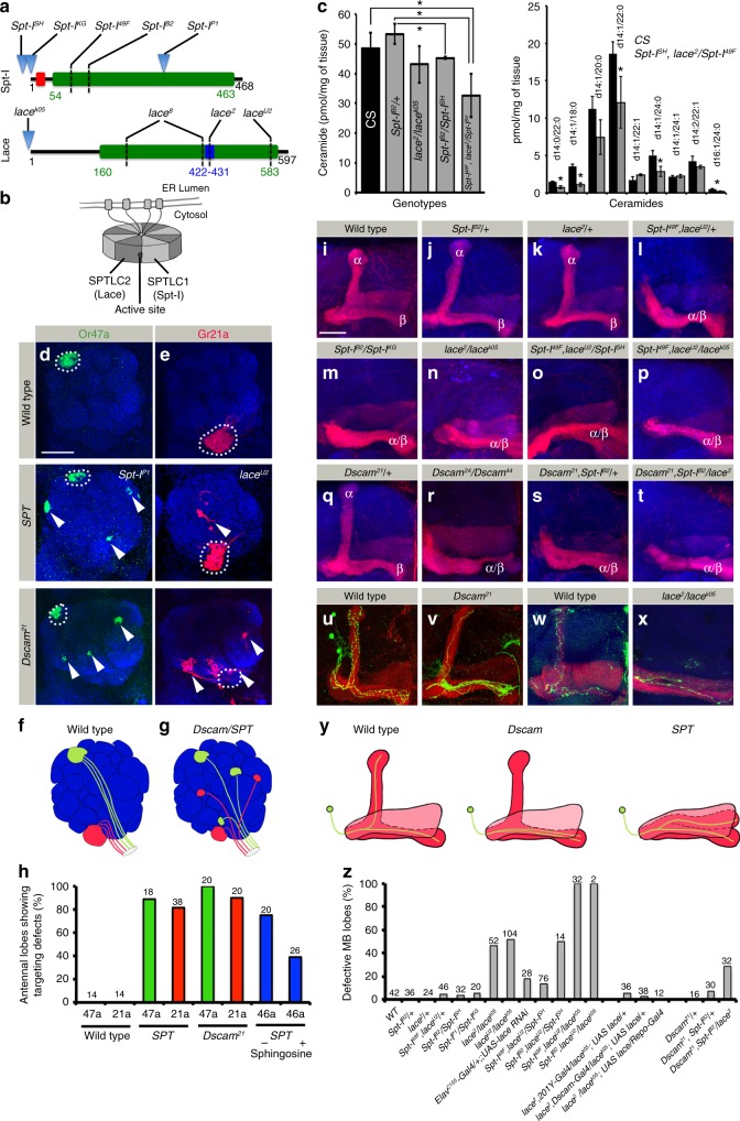
Fig. 1.
Loss of SPT leads to Dscam-like phenotypes in neuronal development. a Protein domain organization of the two SPT subunits of Drosophila, indicating the mutations used in the study. Green: amino-transferase domain, Red: N-terminal transmembrane domain in Spt-I, and Blue: a PLP binding site in Lace. Mutations indicated: Spt-ISH (Spt-ISH1626): P-element insertion in 5’ UTR; Spt-IKG (Spt-IKG06406): P-element insertion at 1st base of SPT-I; Spt-IB2: G127E; Spt-I49F (Spt-I l(2)49Fb4): Q90 stop; Spt-IP1: P-element at Glu295; lace2 (laceHG34): S429N; lace8 (laceVT2): Y221S and K414Q; laceU2: C570T; lacek05 (lacek05305): P-element insertion 8–10 bp upstream of transcription start site. b Schematic showing the octameric organization of SPT holozyme26, 28. Black Dot: Active site. c In adult flies, MS analysis showed that 5 out of 9 identified ceramide species have significantly lower levels in SPT mutants as compared to CS (Right panel), leading to significantly reduced total Ceramide levels (Left panel). Bars represent mean + /− SD across 3 biological replicates. Raw data in Supplementary Data 1. Two sided T-Test *P value < 0.05. d–h Homozygous mutant clones of Spt-IP1, laceU2 and Dscam21 show axonal mistargeting defect (arrowheads) of ORN classes Or47a ((d), green) and Gr21a ((e), red), summarized in the schematics (f, g), and quantified in h. The wild-type targeting site is marked with dotted circle. h In addition, mistargeting of Or46a (blue bars) in lace2/lacek05 is rescued following sphingosine supplementation. i–t Adult MB lobe morphology in Wild type (control) and heterozygous Spt-I and lace mutants show normal α/β lobe segregation (i–k, z) whereas double/trans-heterozygous mutants show defective MB axonal morphology (l–p, z). Dscam and SPT mutants display synergistic effect on MB lobe development (q–t). u–y MARCM clones (Green) of wild type (u) and Dscam21 mutant (v) MB neurons show non-segregated axon branches. Single neuron labeled in wild type (w) and lace trans-heterozygous (x, lace2/lacek05) background using flybow/flip out cassette. y Schematic showing the axonal phenotype in Dscam and SPT mutants in MB of Drosophila. z Quantification of MB lobe defects in different genetic backgrounds. i–x Red: FasII (strongly labels α/β lobes and faintly γ lobe). d, e, i–x Blue: N-Cad (neuropil marker). h, z Numbers on the bars represent number of OL/MB analyzed. Scale Bar: 25 μm

