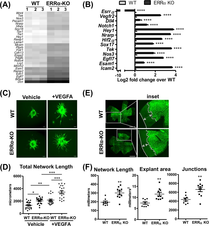
FIG 2.
Loss of ERRα increases angiogenesis in murine lung endothelial cells. (A) Heat map of differentially expressed genes belonging to the GO category “blood vessel morphogenesis” (GO:0048514) from the WT and ERRα-KO cells. (B) Expression of candidate angiogenic genes in ERRα-KO versus WT cells (n = 3). ****, P < 0.00005, unpaired Student's t test. (C) Representative images of calcein AM-stained sprouting angiogenesis in WT and ERRα-KO cells treated with vehicle or VEGFA (30 ng/ml) for 12 h. Scale bars, 100 μm. (D) Quantification of sprouting presented as total network length measured using ImageJ and the Sprout Morphology plug-in (n = 3 experiments, 10 to 20 spheroids per replicate). *, P < 0.05; **, P < 0.005; ***, P = 0.0001, all by Tukey’s multiple-comparison test. (E) Representative images of isolectin B4-stained ERRα-KO P5 mouse retinas and WT littermate controls showing developmental angiogenesis. Scale bars, 1,000 μm. (F) Quantification of explant area, total network area, and number of junctions in retinal vasculature was performed using AngioTool (n = 8 to 10). **, P < 0.005, unpaired Student's t test.

