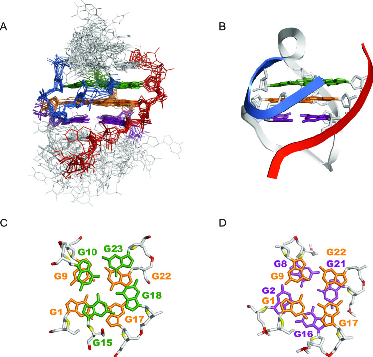
Figure 4.
Solution structure of Otel3Δ2/P6. (A) The 10 lowest energy structures are superimposed. The guanine residues in the top, middle, and bottom G-tetrads are indicated in green, orange, and purple, respectively. The backbone of the V-shaped turn (G15–G18) is indicated in marine. The backbone of the short chain P6 is colored in red. Edgewise loops are colored in light gray. (B) Ribbon representation of a representative refined structure. (C) Stacking of G10-G15-G18-G23 (base in green) over G1–G17–G22–G9 (base in orange). (D) Stacking of G1–G17–G22–G9 (base in orange) over G2–G16–G21–G8 (base in purple). The backbone P is colored in red, and the sugar O4′ is colored in yellow. The other atoms of the backbone are colored in light gray.

