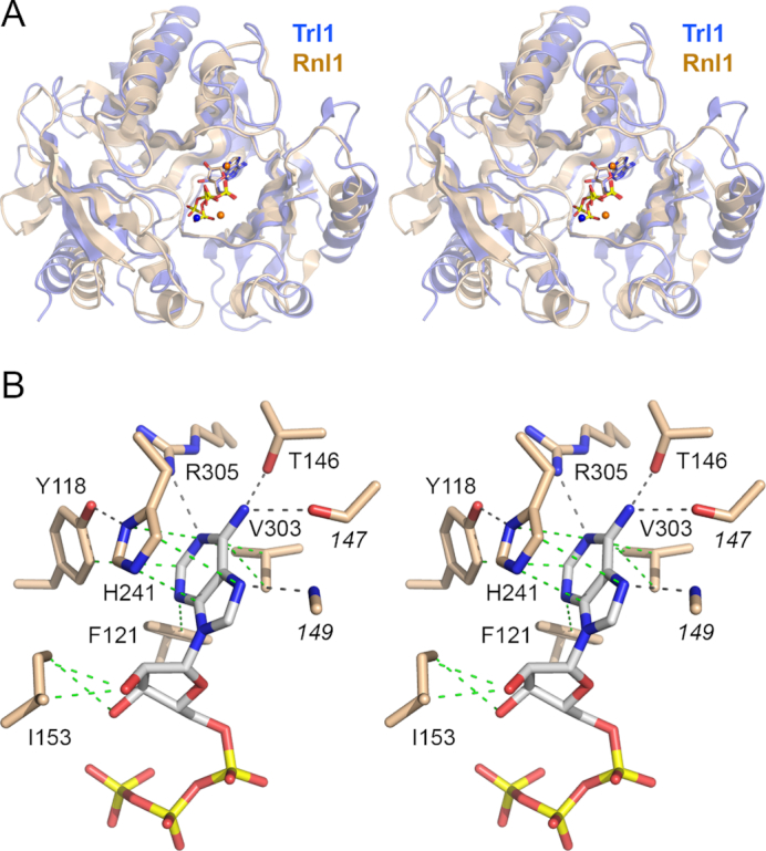
Figure 3.

Homology to T4 Rnl1 and basis for adenine nucleotide specificity. (A) Stereo view of the superimposed structures of the adenylyltransferase domains of Trl1-LIG (blue) and T4 Rnl1 (beige) in their respective complexes with ATP (stick models) and divalent cations (shown as blue spheres for Trl1-LIG and beige spheres for Rnl1). (B) Stereo view of the adenylate binding pocket highlighting contacts to the adenine nucleobase and ribose sugar. Amino acids and ATP are shown as stick models with beige and gray carbons, respectively. Atomic contacts are indicated by black dashed lines (hydrogen bonds) or green dashed lines (van der Waals contacts).
