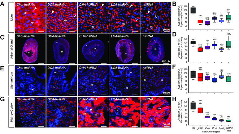
Figure 4.
Distinct cellular uptake and efficacy patterns of lipid-conjugated hsiRNAs. Tissue-dependent internalization of lipid-hsiRNAs 48 h after a single, subcutaneous injection (n = 3 mice, 20 mg kg−1) in (A) liver, (C) adrenal gland, (E) uterine horn and (G) kidney cortex. Cy3-labeled lipid-hsiRNAs (red), nuclei stained with DAPI (blue). Arrowheads described in text. c: cortex; m: medulla; lp: lamina propria; eg: endometrial gland; pct: proximal convoluted tubule; dct: distal convoluted tubule; g: glomerulus. Quantification of Ppib silencing by non-labeled lipid-hsiRNAs in (B) liver, (D) adrenal gland, (F) uterine horn and (H) kidney cortex. Ppib mRNA levels were measured with QuantiGene 2.0 (Affymetrix) assay and normalized to a housekeeping gene, Hprt. All data presented as percent of saline-treated control. All error bars represent mean ± SD. *P < 0.05; **P < 0.01; ***P < 0.001; ****P < 0.0001 as calculated by one-way ANOVA with Tukey's test for multiple comparisons.

