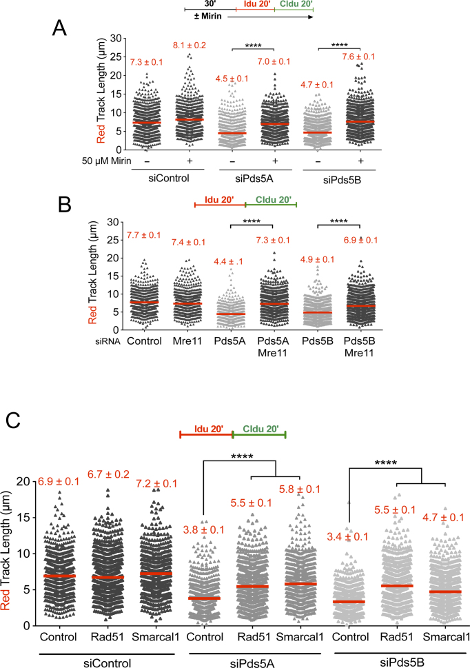
Figure 3.
MRE11 degrades reversed replication forks in the absence of Pds5. (A) Single-molecule DNA fiber labeling scheme (top). Size distribution of IdU tract length in Pds5A and Pds5B siRNA depleted U-2 OS cells treated with 50 μM Mirin. Bars represent the mean. Out of two repeats; n ≥ 300 tracts scored for each data set. Statistics: Mann–Whitney; ****P < 0.0001. (B) Size distribution of IdU tract length in Pds5A, Pds5B, MRE11, Pds5A/MRE11 and Pds5B/MRE11 siRNA depleted U-2 OS cells. Bars represent the mean. Out of two repeats; n ≥ 300 tracts scored for each data set. Statistics: Mann–Whitney; ****P < 0.0001. (C) Size distribution of IdU length in Rad51 or Smarcal1 siRNA depleted U-2 OS cells in the presence and absence of Pds5A or Pds5B. Bars represent the mean. Out of three repeats; n ≥ 300 tracts scored for each data set. Statistics: Mann–Whitney; ****P < 0.0001.

