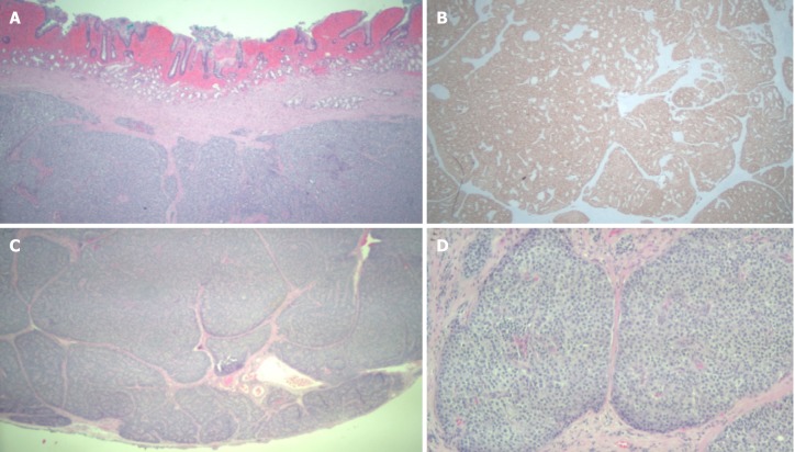
Figure 1.
Pathology of duodenal mass. A: Well differentiated neuroendocrine tumor, hematoxylin and eosin (HE) stain, 25 ×; B: The tumor cells are immunoreactive for synaptophysin (immunohistochemical stain, 25 ×); C: Well differentiated neuroendocrine tumor with negative deep margin (R0, HE stain, 25 ×); D: Well differentiated neuroendocrine tumor (HE, 100 ×).

