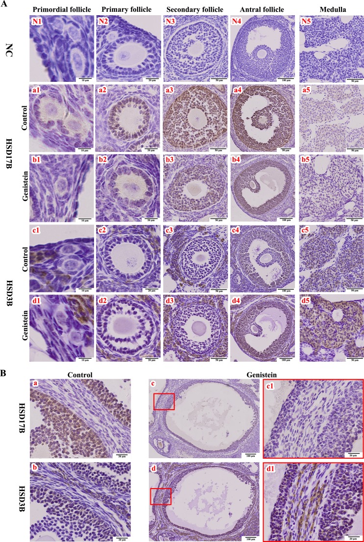
Fig. 5.
Immunohistochemical staining of HSD17B and HSD3B proteins in ovaries. A) HSD17B and HSD3B staining at different stages of follicular development, and in cortex from controls (a1–a5 and c1–c5) and neonatal genistein-treated ovaries (b1–b5 and d1–d5). B) HSD17B and HSD3B staining in cystic follicles from neonatal genistein-treated ovaries (c–c1 and d–d1) and in normal large follicles from control ovaries (a and b). NC (N1–N5), negative control; HSD17B, 17β-hydroxysteroid dehydrogenase; HSD3B, 3β-hydroxysteroid dehydrogenase.

