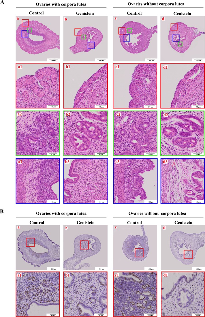
Fig. 6.
Effects of neonatal genistein exposure on uterine morphologic migration and Ki67 expression in mice at PND 90 corresponding to the status of the ovary (with or without corpora lutea). A) Uterine histology. a and c represent the development of uteri from control mice. b and d represent the development of uteri from neonatal genistein-exposed mice. a1–a3, b1–b3, c1–c3, and d1–d3 are the partially magnified drawings of a, b, c, and d, respectively; B) Immunohistochemical staining of Ki67 protein in uteri from control (a–a1 and c–c1) and neonatal genistein-exposed mice (b–b1 and d–d1).

