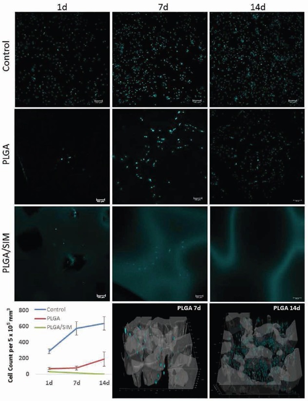
Fig. 4.

Mesenchymal stem cell (MSC) quantification after 1, 7 and 14 days of culture in the scaffolds. Cell nuclei were stained with DAPI (blue) and analyzed by laser scanning confocal microscopy (LSCM). The 3D reconstructions of poly(lactic-co-glycolic acid) (PLGA) pure scaffold merged with adhered cells are presented at the bottom of the image. 3D reconstructions of PLGA/simvastatin (SIM) with cells were not carried out due to SIM autofluorescence, blurring the visualization of the cells. Also, 3D reconstructions of PLGA after 1 day in culture were not performed because there were few cells to justify the reconstruction. The cell count graph represents the median and standard deviation of a triplicate assay.
