Figure 1.
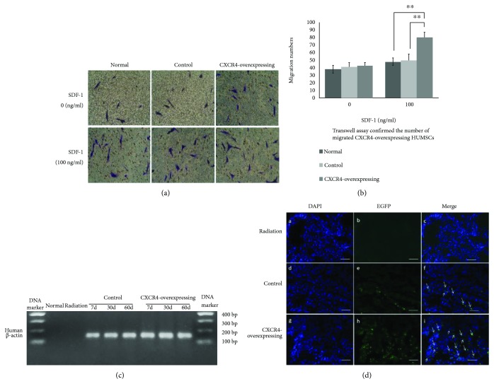
Effect of CXCR4 overexpression on HUMSCs' migration and distribution. (a) The migrated HUMSCs in the down side of the polycarbonate membrane in transwell by hematoxylin staining under white light microscopy at 200x magnification. (b) Transwell assay confirmed that the number of migrated CXCR4-overexpressing HUMSCs was increased compared to control and normal HUMSCs when the under chambers contained SDF-1 (∗∗ P < 0.01, n = 5). There was no difference between every group not containing SDF-1. (c) Examination of human β-actin expression in mouse lung tissues by detecting cDNA with hACTB-specific primers. The expression of β-actin only present in human but not in mouse was detected in control and CXCR4-overexpressing groups and was absent in normal and radiation groups. (d) The EGFP-positive cells were detected by the fluorescence examination in frozen lung sections. The quantity of EGFP-positive cells was much more in the CXCR4-overexpressing group compared to control group (DAPI: blue; EGFP: green).

