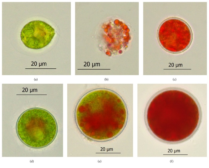
Figure 2.
Photomicrographs of H. pluvialis cells grown in the red stage. Green motile cell (a) and nonmotile cell (d). The motile cell (b) and nonmotile cell (e) after exposure to phosphate and nitrate starvation at 80 μmol photons m−2 s−1 for 3 days. The red cyst formed after 9-day induction in the motile cells cultures (c) and nonmotile cells cultures (f).

