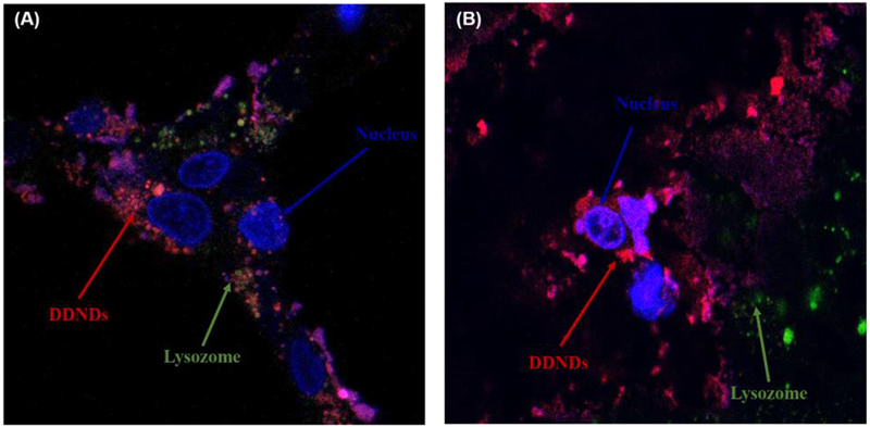
Figure 4.

Confocal images of DDND systems prepared at N/P ratio of 60 using dye attached PB copolymer in (A) Capan-1 and (B) CD18/HPAF cells. Incubation time: 24 h. Red: DDND stained by Alexaflour647, Green: lysosome stained by Lysotracker Green, Blue: nucleus stained by Hoechst dye.
