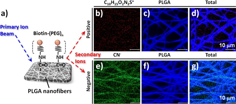
Fig. 4.
a Schematic representation of the conjugation of PEGylated biotin on the surface of the PLGA nanofiber arrays for ToF–SIMS chemical imaging. b–g ToF–SIMS chemical images of PEGylated biotin-conjugated PLGA nanofibers in b–d positive ion mode for b C10H15O2N2S+, c PLGA, and d total ions and e–g negative ion mode for e CN−, f PLGA, and g total ions

