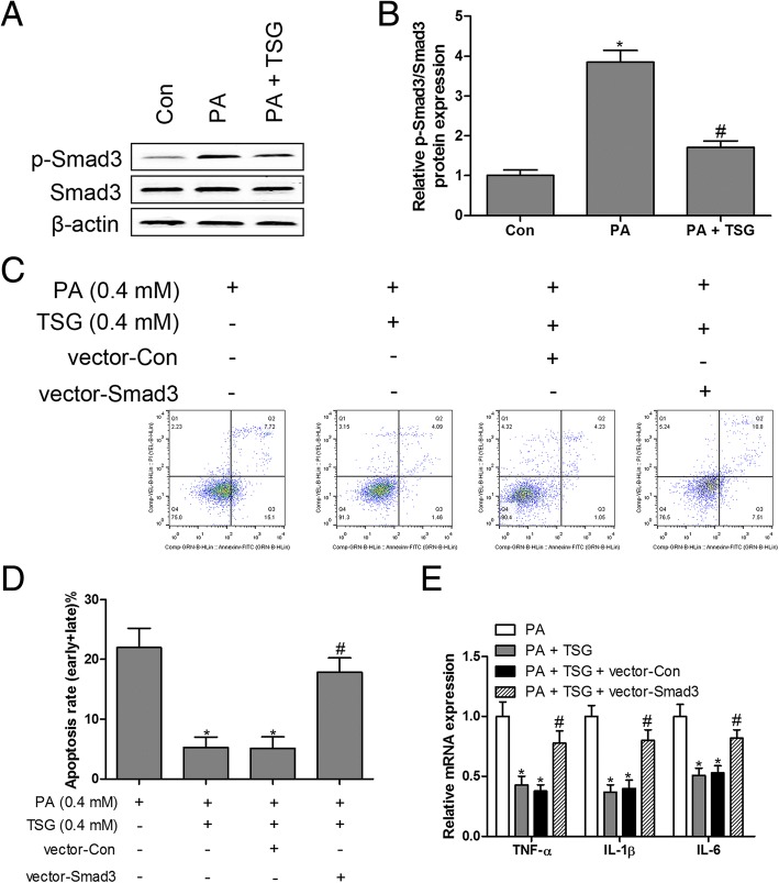
Fig. 3.
Overexpression of Smad3 neutralized the protective effects of TSG in PA-induced inflammation and apoptosis. Protein expression of Smad3 and p-Smad3 was measured by western blotting (a and b). H9c2 cells transfected with Smad3 overexpressed plasmids and treated with PA (0.4 mM) and TSG (0.4 mM) for 48 h, cell apoptosis was detected by flow cytometry (c and d); RT-qPCR was performed to measure the mRNA expression of TNF-α, IL-1β and IL-6 (e). * P < 0.05 compared with control group; # P < 0.05 compared with PA or PA + TSG group. n = 3 in each group

