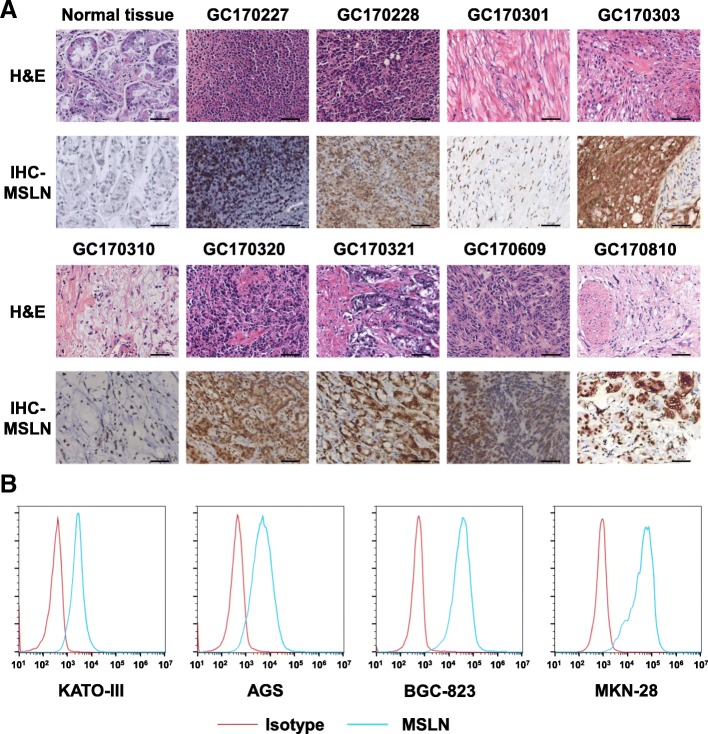
Fig. 1.
MSLN expression in primary GC tissues and cell lines. a Immunohistochemical staining for MSLN in normal gastric tissue and nine primary GC samples, scale bar = 100 μm. b Detection of MSLN expression in four human GC cell lines, including KATO III, AGS, BGC-823, and MKN-28 cells, by flow cytometry

