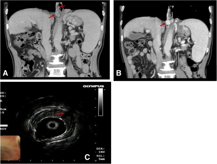
Fig. 3.
Comparison of CT vascular images before and after endoscopic treatment. a: Before the endoscopic treatment, varicose veins and esophageal varices near the esophagus can be seen. b: After endoscopic treatment, esophageal varices disappear, and varicose veins around the esophagus still exist. It is not well detected the perforating veins (PFV),but EUS can be shown(c:arrow)

