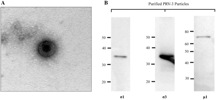
Figure 1.
Purified PRV-3 particles. A Transmission electron microscopy (TEM) of purified PRV-3 viral particles. B Western blot analysis of proteins from purified PRV-3 particles using antiserum raised against PRV-1 σ1, σ3 and µ1 proteins. Observed bands correspond to predicted full length proteins of 35 kDa, 37 kDA and 74 kDa, respectively.

