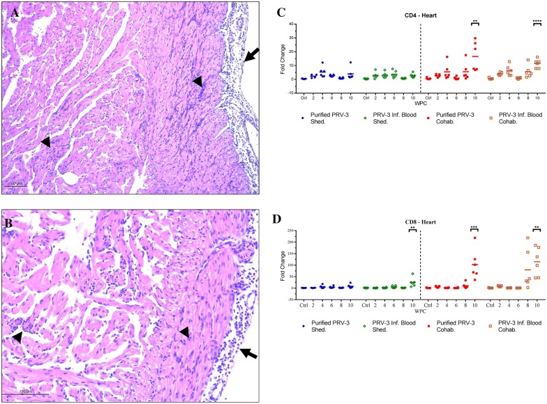
Figure 3.
Histopathological findings and T-cell markers in the hearts of PRV-3 infected fish. A, B Rainbow trout infected with PRV-3 purified particles Epicarditis (long arrows) and inflammation in stratum compactum (outer layer) and stratum spongiosum (internal layer) of heart ventricle (arrow heads) (H&E). C Relative expression of the T-cell marker CD4 in the heart. The Ct values were normalized against control fish from 0 wpc. Significant differences compared to control are shown in asterisks *P < 0.05 **P < 0.001 and ***P < 0.0001. D Relative expression of the T-cell marker CD8 in the heart. The Ct values were normalized against control fish from 0 wpc. Significant differences compared to control are shown in asterisks *P <0.05 **P <0.001 and ***P <0.0001.

