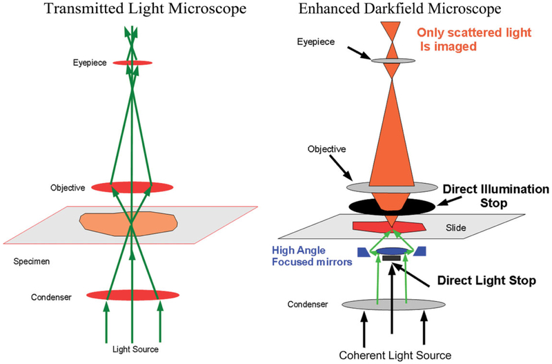
Figure 1.
Comparison of light paths in traditional transmission light microscope and enhanced dark-field microscopy (EDM). The green arrows in the image of the left figure illustrate the light path for a traditional transmission light microscope. In the optics of the EDM, illustrated on the right side of the image, the substage light source is reflected by a cardioid annular condenser mirror to focus the illumination at a high angle of incidence on the tissue section. At this high angle of incidence, transmitted light passing through the tissue section is blocked by the direct illumination stop. The light reaching the eyepiece or camera is restricted. Only light which is scattered due to passage through a submicron particle, whose refractive index differs significantly from the refractive index of the tissue section, will be imaged.

