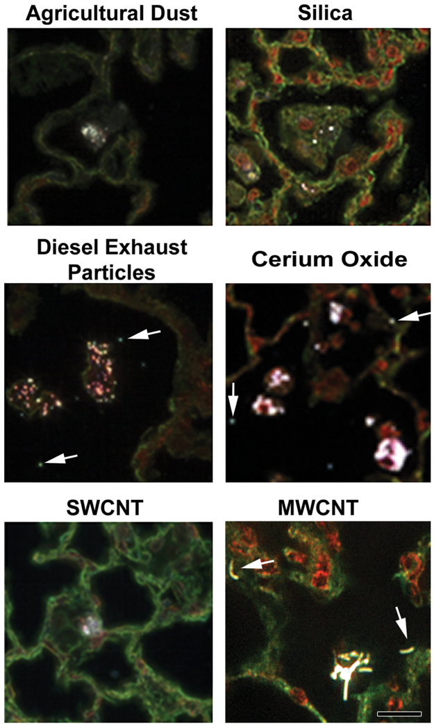
Figure 6.
Examples of particles in the lungs imaged by enhanced dark-field microscopy (EDM). The top row shows pulmonary deposition from typical micron-sized particles (agricultural dust and silica). The second row shows the example images of diesel exhaust particles and cerium oxide (a nanoparticle used as a diesel fuel catalyst). The bottom row shows the images of single-wall carbon nanotube and multiwalled carbon nanotube (MWCNT). Arrows indicate particles outside of alveolar macrophages in the lungs (diesel exhaust particle, cerium oxide, and MWCNT). Particles are white in these EDM micrographs. Cell nuclei in these micrographs are red to brown, while nonnuclear tissue is green in these picrosirius and hematoxylin-stained sections. Calibration marker is 20 μm.

