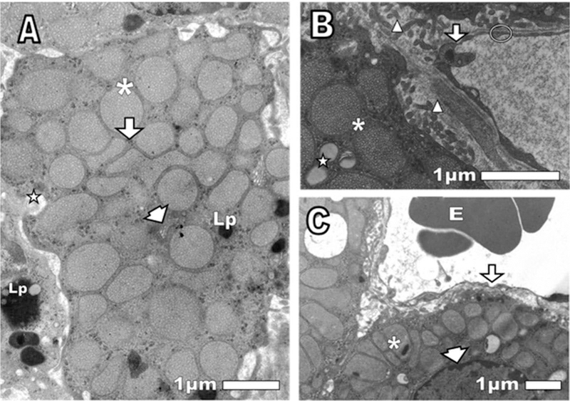
Figure 3.

(A, B, and C) Normal control cortex adrenal gland. After 24 of saline intravenously injection was observed (A) asterisk: mitochondria with tubular cristae. Arrowhead: Golgi apparatus. Arrow: smooth endoplasmic reticulum. Lp: lipofuscin granules in the central cell and a neighboring cell. Star: partially extracted lipid droplet. (B): Triangle: microvilli of cortical cells (way to increase the secretory activity). Arrow: endothelial fenestrae, thin endothelial wall not thickened, as befits a normal capillary. Circle: thin basal membrane. (C): Asterisk: mitochondria with osmiophilics granules. Arrow: endothelium fenestrae not thickened. E: erythrocyte. Arrowhead: the narrow space nuclear envelope, as suitable for a normal cell.
