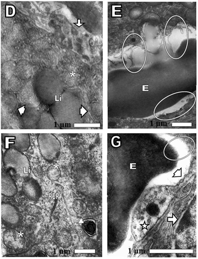
Figure 4.

(D, E, F, and G) Pathological changes in cortex adrenal gland. After 24 of CRP intravenously injection was observed (D) Li: lipid drops, slightly osmiophilics or electrondense. Arrow: continuity of capillary endothelial wall was lost. Arrowhead: smooth endoplasmic reticulum (SER). Asterisk: mitochondria (E) oval: loss of the endothelium and the plasma membrane of cortical cell. On the lower oval, only the cortical cell surface without the plasma membrane was observed. E: erythrocyte. (F) Asterisk: mitochondria swollen and tubular cristae loss. SER was not observed. Triangle: autophagic vacuole. Li: lipid droplets. RER: rough endoplasmic reticulum. (G) Arrow head: fenestrae. Oval: disappearance of the endothelial wall and the surface of the cortical cell without plasma membrane. E: erythrocyte. Star: note the absence of caveolae and pinocytotic vesicles. Arrow: nuclear envelope of cortical cell showing the outer and inner membranes enclosing the perinuclear cistern and a connection site, where the outer membranes of the nuclear envelope looks swollen and is continuous with those of the rough endoplasmic reticulum.
