
Figure 1.
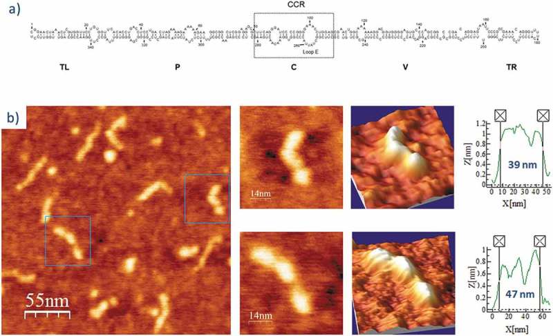
(A) Predicted secondary structure of PSTVd-ml(+). The 359 nt-long, rod-like secondary structure of PSTVd shows the five domains characteristic of members of the family Pospiviroidae: Terminal Left (TL), Pathogenic (P), Central (C), Variable (V), and Terminal Right (TR) [77]. The Central Conserved Region (CCR) is located within the C domain and contains an UV-sensitive loop E motif stabilized by non-canonical base-pairs. Adapted from [26]. (B) AFM images of PSTVd-ml(+), renatured in the absence of Mg2+. A field of 275 × 275 nm is shown on the left panel and two characteristic molecules are zoomed on the right one. 3D views of the imaged viroid RNAs, as well as profiles with their measured lengths (X [nm]) and heights [Z [nm]), are also displayed.