
Figure 9.
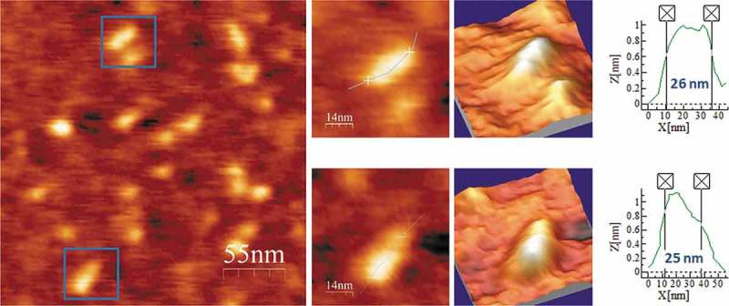
AFM images of ELVd-ml(+) renatured in 4 mM Mg2+. A field of 275 × 275 nm is shown on the left panel and two characteristic molecules are zoomed on the right. 3D views of the imaged viroid RNAs, as well as profiles with their measured lengths are also displayed.