
Figure 11.
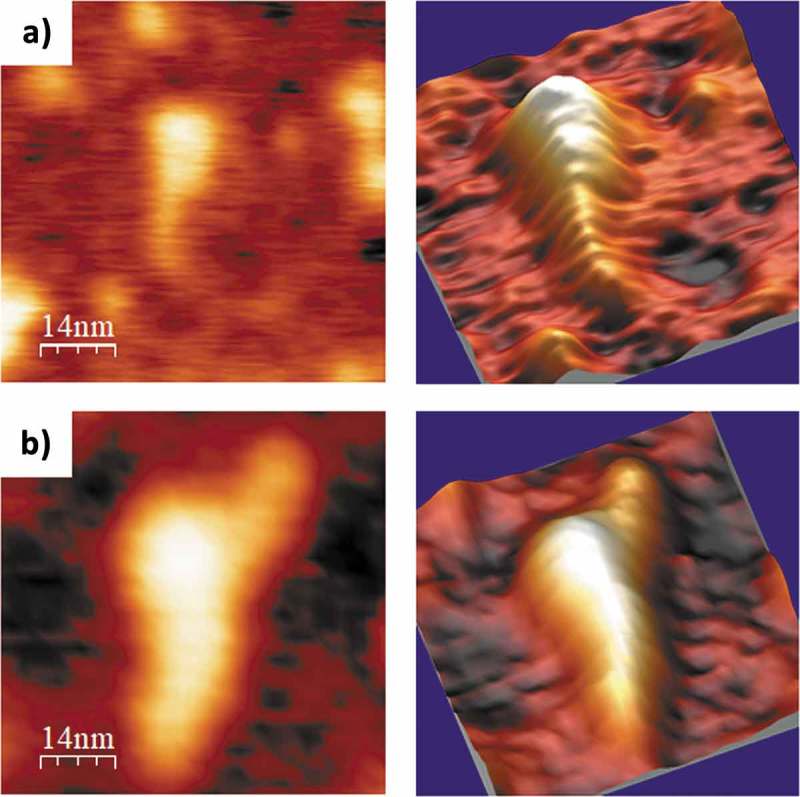
AFM images of selected PLMVd-ml(+)wt (A) and PLMVd-ml(+)mut (B) molecules, renatured in 4 mM Mg2+. 2D (left) and 3D (right) views are displayed, which clearly show the difference in compactness among the wild type (see secondary structure in Fig. 5A) and mutant (see secondary structure in Fig. 7A) viroid RNAs, as well as the presence of a short and flat arm protruding from the head of the latter.