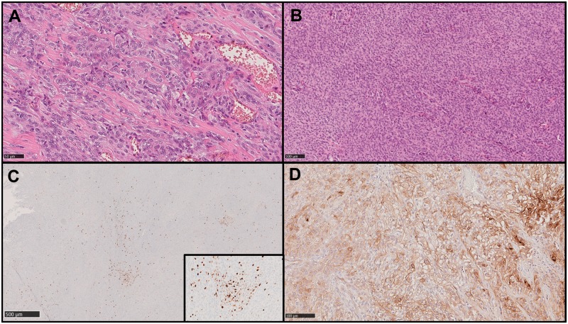
FIGURE 1.
Example images showing some of the morphological and immunohistochemical features assessed. Hematoxylin and eosin (H&E) images showing diffuse prominent nucleoli (A) and sheeting (B). Original magnification: ×200. Immunohistochemical images showing Ki67 hot spots (C) and only partial staining of tumor cells for SSTR2a (D) in a meningioma which recurred during follow up.

