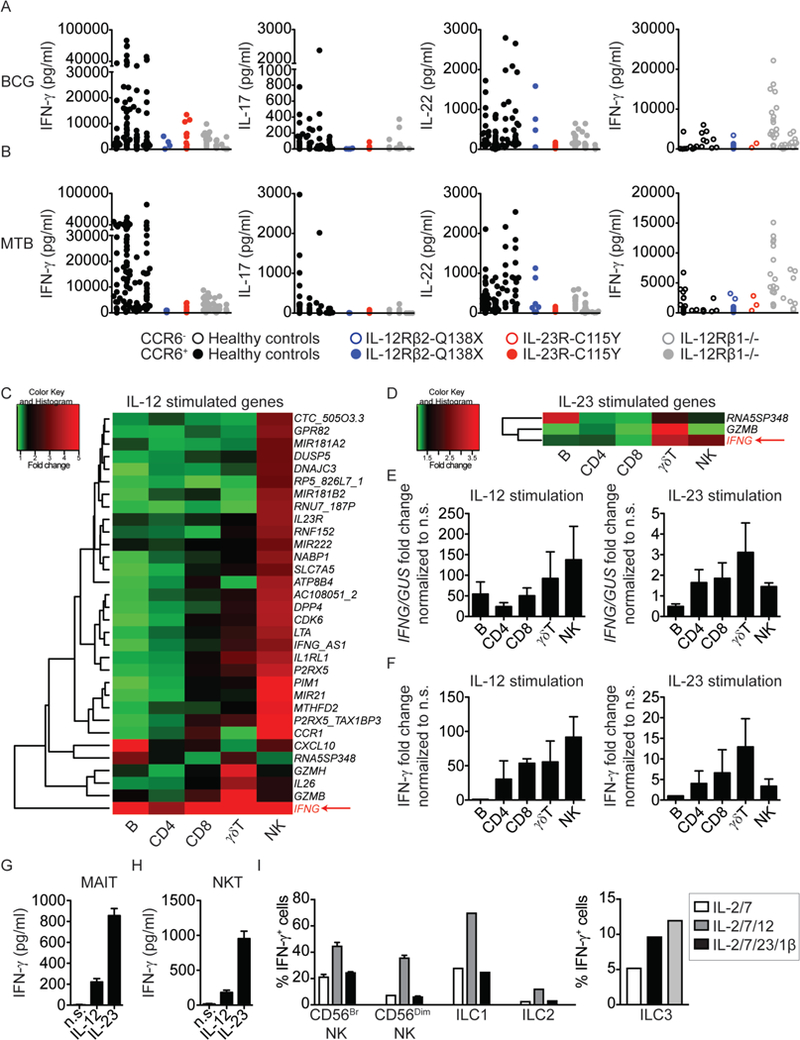
Figure 4: Specific defects in the IL-12- and IL-23-dependent generation of an IFN-γ immune response in patients with novel homozygous mutations of IL12RB2 or IL23R.

A, B) Multiple CCR6+ or CCR6− memory CD4+ T-cell lines were generated by the polyclonal stimulation of sorted peripheral blood subsets from healthy controls (n=4), one IL-12Rβ2-Q138X patient, one IL-23R-C115Y patient, and three IL-12Rβ1-deficient patients. Lines were screened for reactivity with peptide pools covering antigens from BCG (upper panel) and MTB (lower panel). BCG- and MTB-reactive CD4+CCR6+ T-cell lines from each individual were selected and the cytokines accumulating in the culture supernatant were determined with a Luminex machine. Each dot on the graph corresponds to a value for a single antigen-reactive T-cell line. CCR6+ T-cell lines are shown as closed circles, and CCR6− cell lines are shown as open circles. C, D) Microarray heat map of isolated B, CD4+ T, CD8+ T, γδ+ T and NK cells stimulated with IL-12 (C) or IL-23 (D) for 6 h. The data shown are the fold-induction relative to non-stimulated (n.s.) cells. The most commonly upregulated gene, IFNG, is highlighted in the lower right corner of each heat map. E) IFNG induction by isolated B, CD4+ T, CD8+ T, γδT, and NK cells from 5 healthy controls, upon stimulation with IL-12 or IL-23 for 6 h, was assessed by qPCR, and the data were normalized relative to n.s. cells. F) IFN-γ levels in the supernatants from the cells used in (E) were analyzed by ELISA and represented as a fold-change, relative to n.s. cells. G, H) Sorted MAIT cells (G) or NKT cells (H) (>95% pure) were left unstimulated, or stimulated with rhIL-12 (20 ng/mL) or rhIL-23 (100 ng/mL) for 6 h. Cell culture supernatants were harvested and used for IFN-γ determination in a multiplex cytokine assay. I) NK cells, ILC1, or ILC2 were sorted, by FACS, from blood samples from healthy donors and cultured in the presence of the indicated cytokines for 24 h. Total ILCs were gated on viable CD45+Lin− (CD3−CD4−CD5−TCRαβ−TCRγδ−CD14−CD19−) CD7+ cells. NK cells were identified as CD56bright and CD56dim, ILC2 as CD56−CD127+CRTh2+ and ILC1 as CD56−CD127+CD117−CRTh2−. IFN-γ levels were determined by intracellular staining. ILC3 were sorted by FACS from the tonsillar tissues of healthy donors, by gating on viable CD45+ Lin−CD7+CD117+NKp44+ cells. ILC3 were cultured for 4 days in the presence of the indicated cytokines, and IFN-γ was then determined by intracellular staining.
