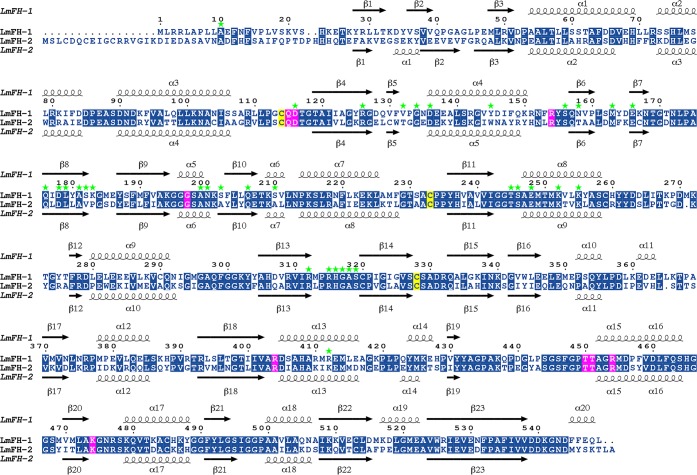
Figure 3.
Sequence alignment of LmFH isoforms. LmFH-1 and LmFH-2 are the mitochondrial and cytosolic isoforms of Leishmania major, respectively. The conserved residues are indicated in the blue boxes. The conserved active site residues among class I FHs that coordinate to inhibitor S-2-thiomalate are indicated in the pink boxes. The three conserved cysteine residues, which are shown to bind a [4Fe-4S] cluster, are indicated in the yellow boxes. Secondary structures of LmFH-1 and LmFH-2 are shown on top and at the bottom of sequence alignment, respectively. The dimer interface residues of LmFH-1 are indicated in green stars. The alignment was performed using MULTALIN15 and graphically displayed using ESPript.16

