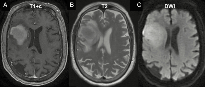
Fig. 1.
PCNSL imaging pattern on MRI. (A) MRI T1 sequence with gadolinium contrast (T1+c) reveals homogeneously enhancing deep lesions. (B) Lesions are iso- to hyperintense on T2 imaging with a relatively small amount of edema. (C) Diffusion-weighted imaging (DWI) demonstrates restricted diffusion in the tumor.

