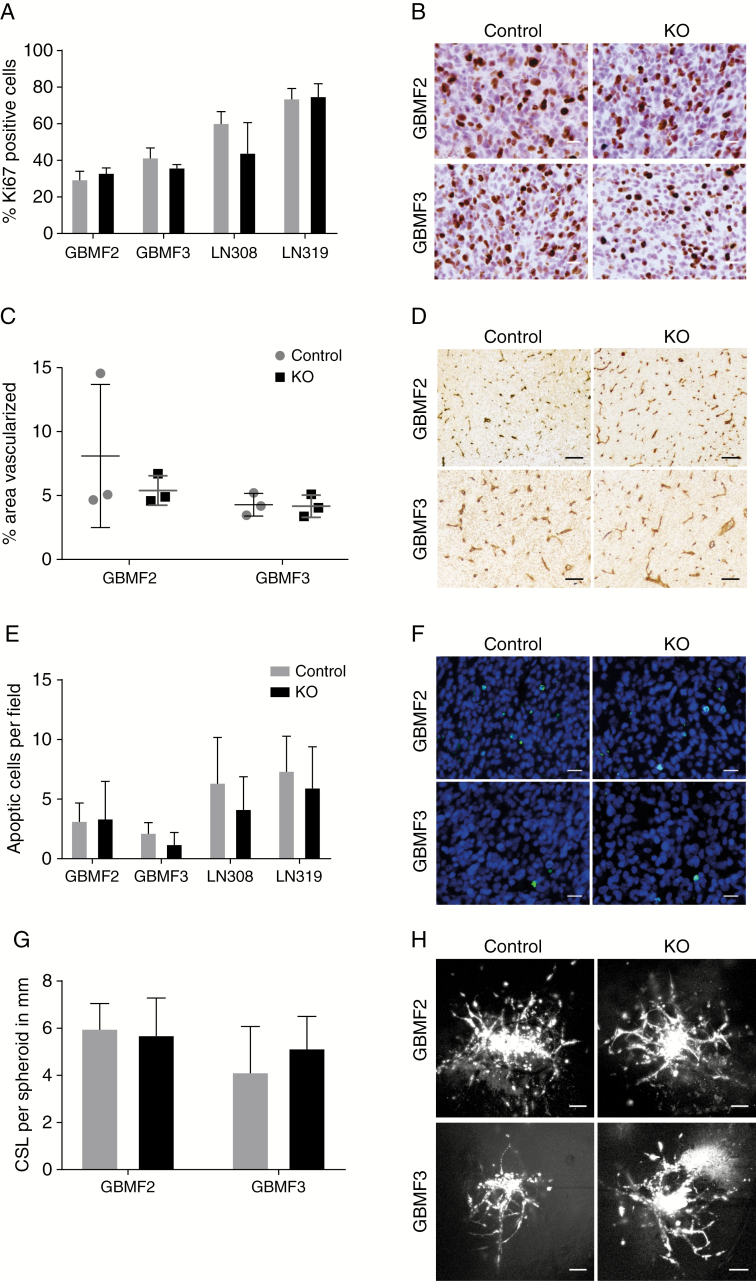
Fig. 4.
The ablation of PDPN has no effect on malignant features. (A) The deletion of PDPN did not influence tumor cell proliferation in vivo as assessed by Ki67 immunohistochemistry (B), 5 fields (0.33 mm2/field) per group analyzed, p(GBMF2) = 0.15; p(GBMF3) = 0.05; p(LN308) = 0.07; p(LN319) = 0.80; Student’s t-test. (C) Tumor area covered by blood vessels (immunohistochemical staining for laminin, D) is not altered between control and PDPNKO groups, 4–5 fields (3.55 mm2/field) of 3 tumors each were analyzed, p(GBMF2) = 0.46; p(GBMF3) = 0.88; Student’s t-test. (E) Quantification of apoptotic cells per field showed no difference between control and knockout tumors, 5 fields (0.33 mm2/field) per group analyzed, p(GBMF2) = 0.90; p(GBMF3) = 0.13; p(LN308) = 0.32; p(LN319) = 0.50; Student’s t-test. (F) Representative pictures of TUNEL staining; apoptotic cells with fragmented DNA are indicated in green. (G) Invasion of PDPNKO and control cells in an ex vivo invasion assay. Quantification of cumulative sprout length (CSL) per spheroid, n(GBMF2) = 12; n(GBMF2KO) = 16; n(GBMF3) = 19; n(GBMF3KO) = 15; p(GBMF2) = 0.60; p(GBMF3) = 0.08; Student’s t-test. (H) Representative pictures of glioblastoma cell invasion. Scale bars (B, F) 20 μm; (D, H) 100 μm.

