
Fig. 1.
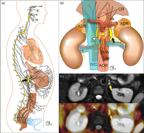
(a) Schematic presentation of the sympathetic system network (black lines) with compact parts called ganglia (oval objects). (b) Schematic presentation of the coeliac ganglia (arrows) location. ADR, adrenal glands; AOR, aorta; CR, crura of the diaphragm; CT, coeliac trunk; IVC, inferior vena cava; SMA, superior mesenteric artery. (c, d) transverse images on the level of celiac ganglia (arrows): (c) MR T2-weighted fat-saturated image. (d) Multimodal 68Ga-PSMA-11 PET/MRI. PSMA, prostate-specific membrane antigen.