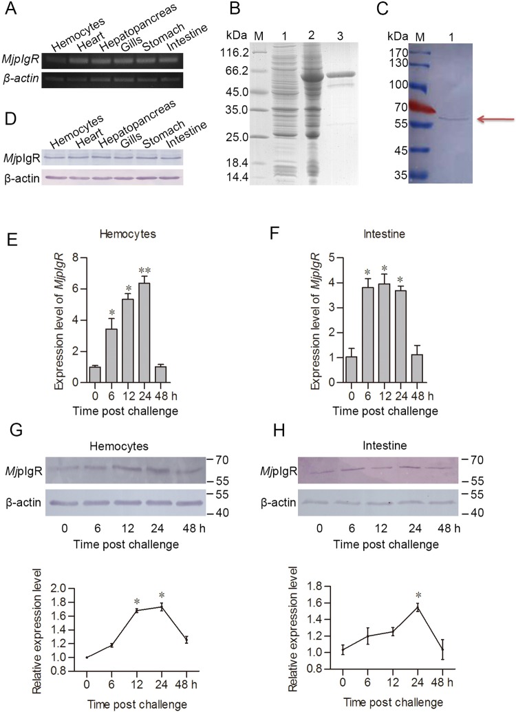
Fig 1. MjpIgR was upregulated in shrimp after WSSV challenge.
A, The tissue distribution of MjpIgR in shrimp at the mRNA level. B, Recombinant expression and purification of the extracellular region of MjpIgR in E. coli. Lane 1, total proteins from E. coli with MjpIgR-pGEX4T-1, without IPTG induction; lane 2, total proteins from the E. coli with IPTG induction; lane 3, purified recombinant MjpIgR; lane M, protein molecular mass marker. C, MjpIgR in normal hemocytes of shrimp was detected using western blotting with MjpIgR polyclonal antibodies. Lane M, protein marker; lane 1, MjpIgR in hemocytes detected using the MjpIgR polyclonal antibodies. D, The tissue distribution of MjpIgR in different tissues was investigated using western blotting. E-F, Expression patterns of MjpIgR in hemocytes (E) and intestine (F), as detected using qPCR. The data were analyzed statistically using Student’s t test. G-H, MjpIgR expression patterns at the protein level in hemocytes (G) and intestine (H) of shrimp after WSSV challenge, as analyzed using western blotting. The lower panels of (G) and (H) show statistical analysis for three replicates. The results are expressed as the mean ± SD. *, p< 0.05; **, p < 0.01.

