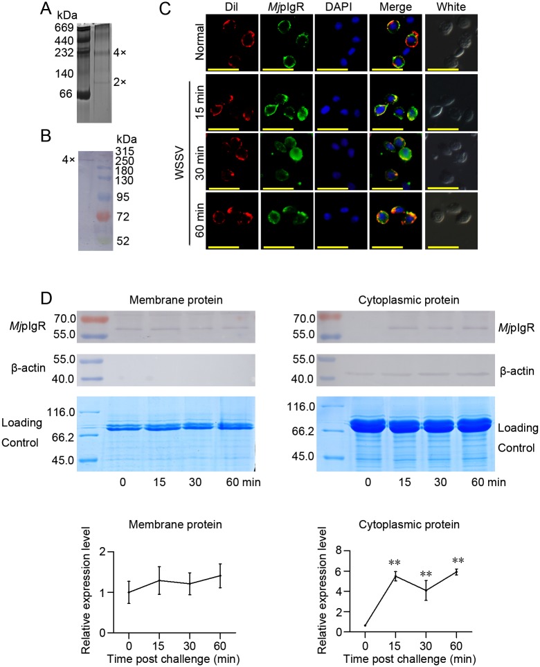
Fig 3. MjpIgR oligomerized to tetramers and internalized into the cytoplasm of hemocytes in shrimp after WSSV infection.
A, Native PAGE of rMjpIgR. Purified rMjpIgR was analyzed using native PAGE and stained with Coomassie blue. B, A tetramer of MjpIgR was detected in vivo using western blotting after treatment of hemocytes with a crosslinker (BS3). Shrimp were injected with WSSV, after 30 min, the hemocytes were collected and treated with BS3. These hemocytes were homogenized and the extracted proteins were separated by SDS-PAGE. Western blotting was then performed using anti-MjpIgR antibodies. C, MjpIgR in hemocytes of shrimp was detected at 0 (untreated), 15, 30, and 60 min post WSSV injection. Scale bar = 20μm. D, Shrimp were challenged with WSSV and then membrane and cytoplasm proteins of the hemocytes were extracted. MjpIgR in the membrane and cytoplasm of hemocytes was analyzed using western blotting at 0, 15, 30, and 60 min post-WSSV injection. The lower panels show the statistical analysis from three independent experiments. **, p < 0.01.

