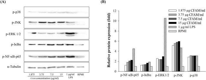
Fig 5. Effects of CFAMs on the proteins associated with NF-κB and MAPK pathways in RAW264.7 cells.
Cells were stimulated with various concentrations of CFAMs or 1 μg/mL of LPS for 24 h. Total protein was extracted from each treatment and was separated on SDS-PAGE. The separated protein was transferred on to PVDF membrane. The membrane was incubated with the specific antibodies and was detected using the Pierce® ECL Plus Western Blotting Substrate and were imaged using the ChemiDoc XRS+ imaging system. (A) Western blots showing protein bands, (B) Relative band intensity.

