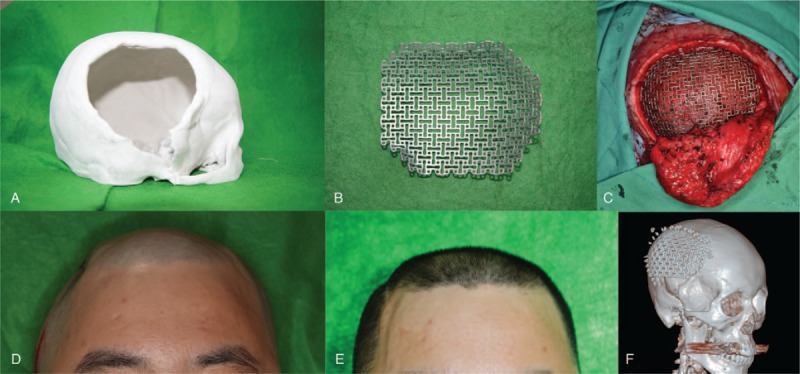
Figure 4.

A 3D printed plaster cast (A) and titanium mesh of the patient (B) were prepared. The titanium mesh was inserted and fixed to the adjacent bone (C). Compared to the preoperative photograph that showed a depressed deformity on the right temporal area (D), after 12 days, the deformity was resolved, and both temporal areas showed symmetry (E). Postoperative 3D-CT reconstruction shows a well-positioned mesh (F). 3D-CT = 3-dimensional computed tomography.
