. 2019 Feb 19;8:e40093. doi: 10.7554/eLife.40093

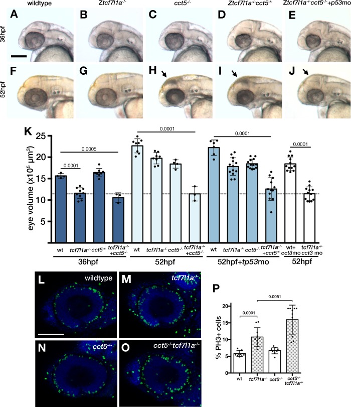
Figure 8. Loss of tcf7l1a modifies the cct5u762 mutant eye phenotype.

(A–J) Lateral views of wildtype (A, F), Ztcf7l1a-/- mutant (B, G) cct5U762/u762 mutants (C, H), double cct5U762/u762/Ztcf7l1a-/- mutants (D, I) and double cct5U762/u762/Ztcf7l1a-/- mutants injected with 0.8 pmol of cct3 morpholino (E, J) at indicated stages. Scale bar = 100 µm. Full data in Supplementary file 10O, single experiment, 36hpf, wt n = 4, Ztcf7l1a-/- n = 9, cct5-/- n = 8, cct5/Ztcf7l1a-/- n = 3; 52hpf, wt n = 8, Ztcf7l1a-/- n = 8, cct5-/- n = 4, cct5/Ztcf7l1a-/- n = 3; 52hpf + 2 pmol tp53 morpholino, wt n = 6, Ztcf7l1a-/- n = 13, cct5-/- n = 13, cct5/Ztcf7l1a-/- n = 12; 52hpf + 0.8 pmol cct3 morpholino, wt n = 12, Ztcf7l1a-/- n = 12. (K) Eye volume quantification at the indicated timepoints and conditions shown in A–J) (data in Supplementary file 1O). Unpaired t-test. (L–O) Immunostaining detecting phosphohistone3 (PH3, green) in wildtype (L), Ztcf7l1a-/- (M), cct5-/- (N), cct5-/-/Ztcf7l1a-/- (O) eyes at 32hpf. (P) Plot showing the percentage of PH3 positive cells in the eyes shown in L–O) (data in Supplementary file 1Q) Single experiment, wildtype n = 10, Ztcf7l1a-/- n = 10, cct5-/- n = 9, cct5/Ztcf7l1a-/-n = 10, unpaired t-tests.

Figure 8.

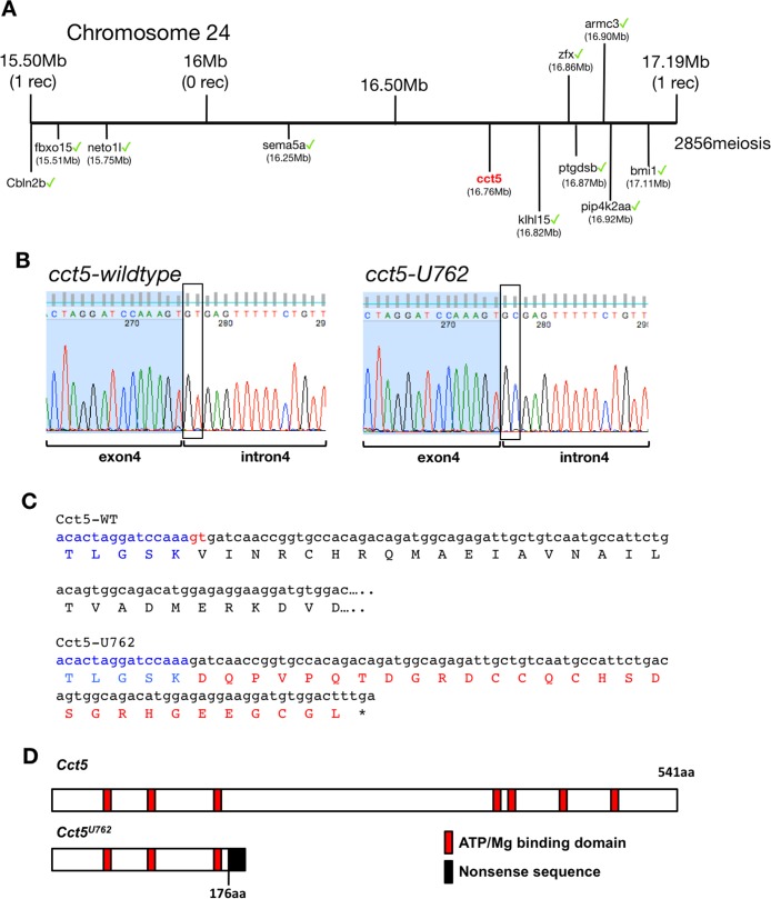
Figure 8—figure supplement 1. Genetic mapping of U762 and description of the cct5U762 mutation.

Figure 8—figure supplement 1.

(A) Representation of the SSLP segregation linkage analysis mapping of U762) modifier of tcf7l1a to a 1.69 Mb interval on chromosome 12, between 15.50 Megabases (Mb) with one recombinant (rec) and 17.19 Mb with one rec. Green ticks highlight sequenced genes in the interval that show no mutations. (B) DNA sequencing chromatograms of the genomic fragment encompassing the 3’ end of cct5 exon 4 and 5’ end of intron four from wildtype (left) and cct5U762/762(right) embryos. Boxes show the splice donor nucleotides in intron 4. (C) Nucleotide and protein sequence of wildtype (top alignment) and cct5U762 (bottom alignment). cct5 exon four nucleotides and amino acids in blue. The last two 3’ nucleotides in exon four that are used in cct5U762 as a splice donor in red. Nonsense amino acid sequence in cct5U762 in red. (D) Cartoon of wildtype and Cct5U762 protein product. Red boxes show Mg2+/ATP binding domains, black box indicates nonsense mutant protein stretch.
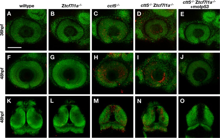
Figure 8—figure supplement 2. Whole mount Tunel cell death analysis in cct5/tcf7l1a mutants.

Figure 8—figure supplement 2.

Tunel assay labelling detecting apoptotic cells (red) performed in wildtype (A, F, K, 100% n = 8), Ztcf7l1a-/- (B, G, L, 100% n = 10), cct5-/- (C, H, M, 100% n = 9), cct5/Ztcf7l1a-/- (D, I, N, 100% n = 4) and cct5/Ztcf7l1a-/- + 2 pmol of tp53 morpholino (E, J, O, 100% n = 5). DAPI pseudocoloured in green. Scale bar = 100 µm.
Figure 8—figure supplement 3. atoh7 expression in cct5/tcf7l1a mutants.

Figure 8—figure supplement 3.

(A–C) Anti-GFP immunocytochemistry (green) showing Tg(atoh7:GFP)- labelled neurons in wildtype. (A) cct5-/- (cct5U762/U762) (B) and cct5-/-/Ztcf7l1a-/- (C) embryos at 48hpf. Dotted line in (C) outlines the eye profile. Arrowhead points to the retina ganglion cell layer. Scale bar = 100 µm.