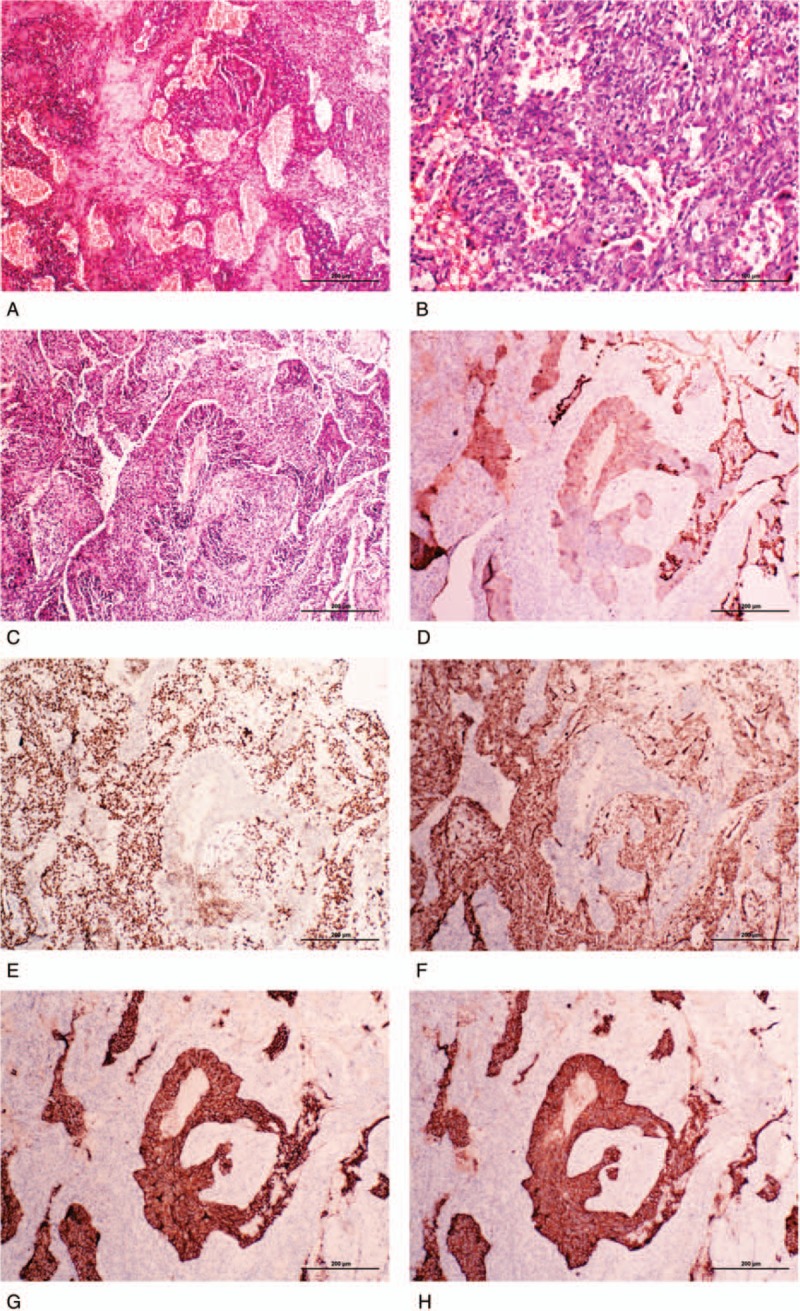
Figure 2.

The histological and immunohistochemical features of the present sclerosing pneumocytoma accompanied with typical carcinoid tumor. (A) Typical hemorrhagic and sclerotic regions of the sclerosing pneumocytoma region of the present sclerosing pneumocytoma. (B) Two cell types were observed in the papillary and glandular region of sclerosing pneumocytoma: surface cuboidal cells and stromal round or polygonal cells. (C) A representative field of sclerosing pneumocytoma mixed with carcinoid tumor. The well-arranged cell cords in the center were carcinoid tumor, and periphery papillary and solid regions were sclerosing pneumocytoma. (D) Immunohistochemical staining of broad-spectrum cytokeratin was strongly positive in the cuboidal cells of sclerosing pneumocytoma and weakly positive in carcinoid tumor cells, but negative in the stromal polygonal cells. (E) Immunohistochemical staining of thyroid transcription factor-1 (TTF-1) was positive in both cuboidal surface cells and stromal polygonal cells, but negative in the carcinoid tumor tissue. (F) Immunohistochemical staining of vimentin was positive in the sclerosing pneumocytoma tissue, but negative in the carcinoid tumor tissue. (G) and (H) Immunohistochemical staining of CD56 and synaptophysin were both positive in the carcinoid tumor tissue, but negative in the sclerosing pneumocytoma tissue.
