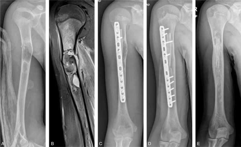
Figure 1.

A 12-year-old male patient with monostotic fibrous dysplasia in the diaphysis of right humerus. (A) X-ray shows the fibrous dysplasia and pathological fractures occur at the diaphysis of right humerus. (B) Magnetic resonance imaging shows T1 hypointense signal at the diaphysis of right humerus, demonstrating the extent of disease. (C) X-ray anteroposterior view on the day of surgery. (D) X-ray at 12 months after surgery showing union. (E) Good healing of the fracture and bony union between the cortical strut allograft and the host bone at 5 years after surgery.
