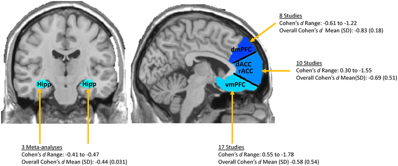
Figure 2. Estimated Effect Sizes of Hippocampal and mPFC Volume Reductions in MDD.
The medial prefrontal cortex (mPFC) includes ventral portions of the mPFC (ventral medial prefrontal cortex; vmPFC, medial orbitofrontal cortex; mOFC) and dorsal portions of the mPFC (dorsal medial prefrontal cortex; dmPFC) as well as rostral anterior cingulate cortex; rACC and dorsal anterior cingulate cortex; dACC). In this figure, we provide a range, mean (standard deviation) of effect sizes calculated from individual studies for each of the mPFC subdivisions. We also provide a range, mean (standard deviation) of effect sizes for reduced hippocampal volumes in MDD calculated from three prior meta-analyses. Negative effect size values indicate that those with MDD have reduced mPFC and hippocampal volumes compared to healthy controls.

