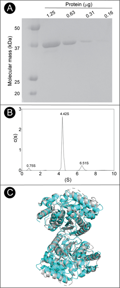
Figure 11. Oligomeric state and purity of MyOobT.

A. SDS-PAGE behavior of MjCobT compared to BioRad Precision Plus Protein Standards. Based on the data shown in panel B, MjCobT was estimated to be >99% homogeneous. B. Sedimentation velocity c(s) distribution of MjCobT at pH7.5 showing three species at 0.75 S, 4.42 S, and 6.51 S C. Cartoon model of the MjCobT dimer (cyan) identified in the crystal structure (PDB ID 3L0Z) superimposed onto the homologous CobT from Pyrococcus horikoshii dimer in grey (PDB ID 3U4G).
